Research Highlights


Welcome to my research page. Below are some of my primary research focuses, including optical microscopy techniques, Laue microdiffraction, and advanced X-ray scattering analysis methods.
DInM Microscope
The DInM microscope is a reflected-light, common-path interferometry technique designed for real-time surface characterization. With an axial resolution of around 0.57 nm, it has been successfully applied to measure step heights, observe in-situ silicon buckling on elastomers, and investigate martensitic phase transformations.
I developed a specialized DInM microscope that can vary the beam shear distance to study specimen surface morphology. For further details, refer to this paper.


Synchrotron Laue Microdiffraction

Laue microdiffraction is widely employed to obtain crystallographic information, grain orientations, localized strains, heterogeneous phases, and precipitates in materials. However, the analysis of Laue microdiffraction data can be both time-consuming and computationally intensive.
The newly developed CANON algorithm generates feature maps of Laue scans without the need for indexing. Additionally, I have implemented a physics-informed approach to enhance the resolution of Laue scans, enabling nano-crystal grain mapping with a micron-sized probe. A supervised machine learning algorithm can index multi-grain data within minutes.


Automatic SAXS, WAXS, GISAXS, GIWAXS Analysis
X-ray scattering extends traditional X-ray diffraction techniques. Many methods used in X-ray diffraction are adapted to analyze scattering data. I have developed scripts to automatically subtract the background and fit the corresponding peaks—a crucial tool for upgraded beamlines with higher flux and more data.

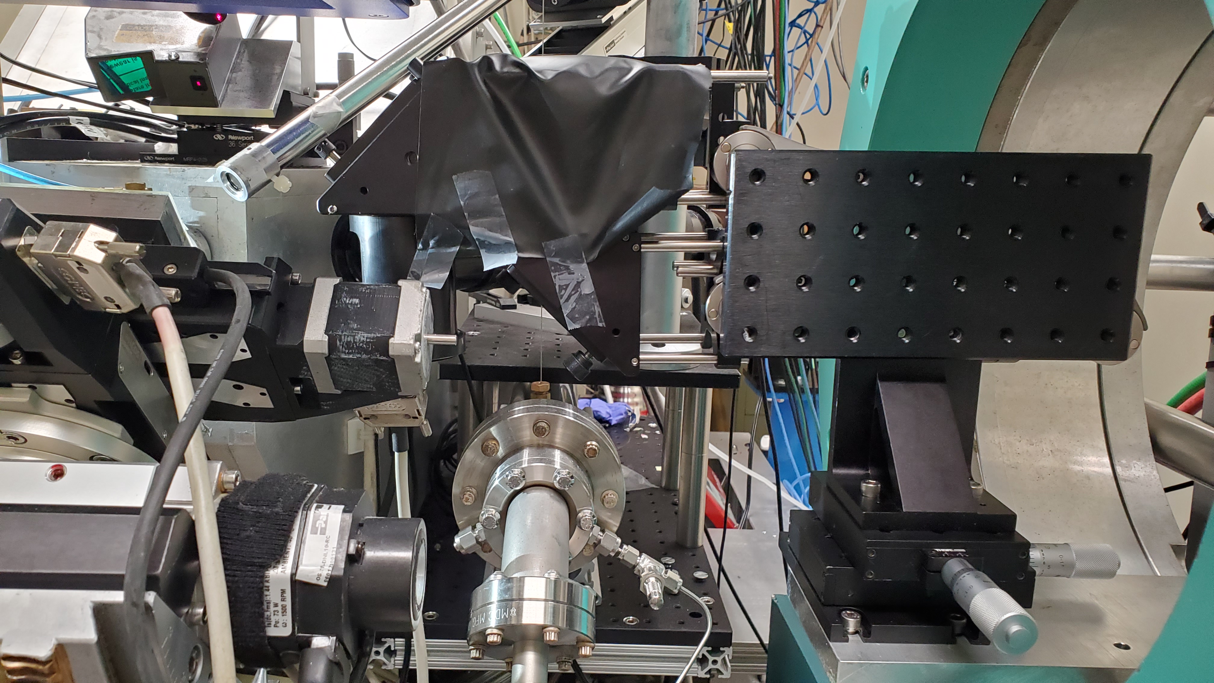
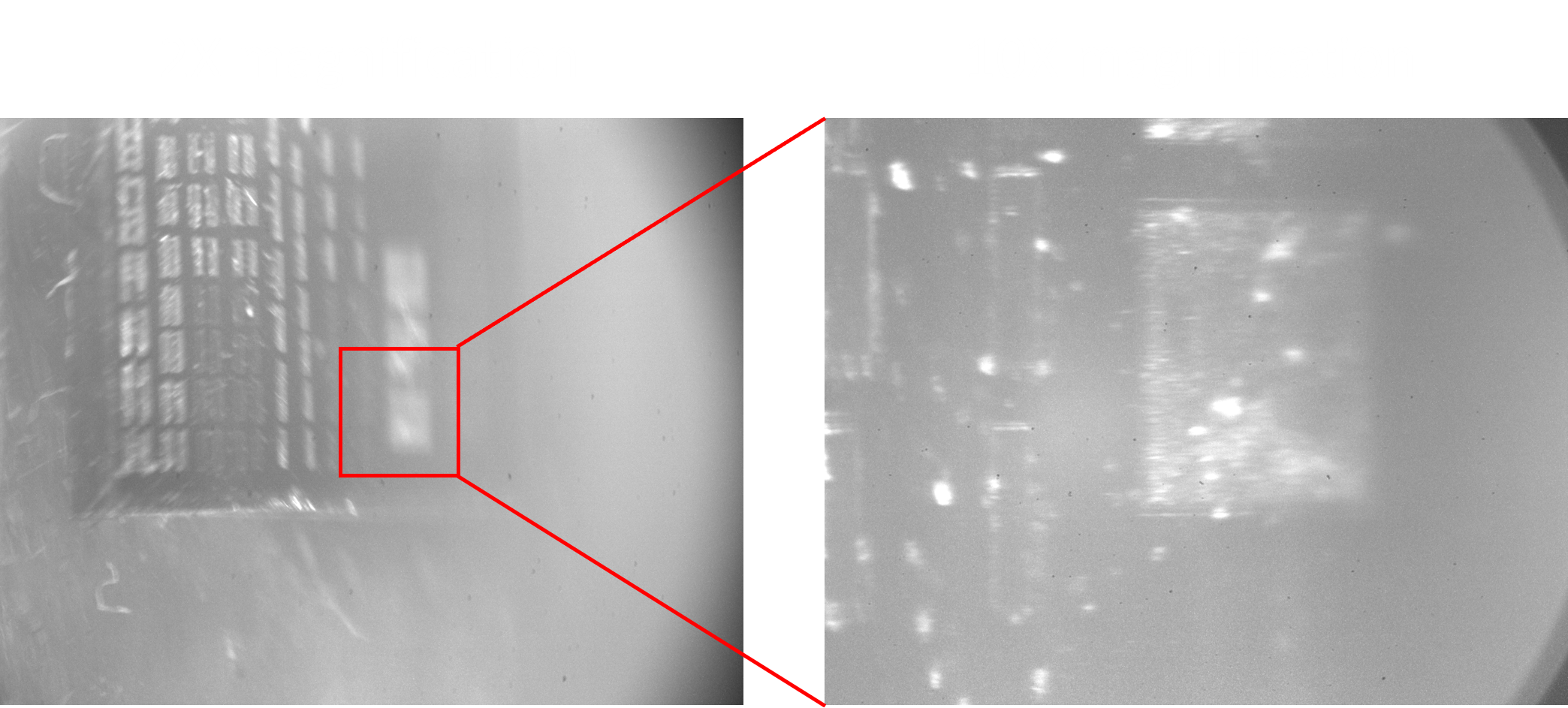
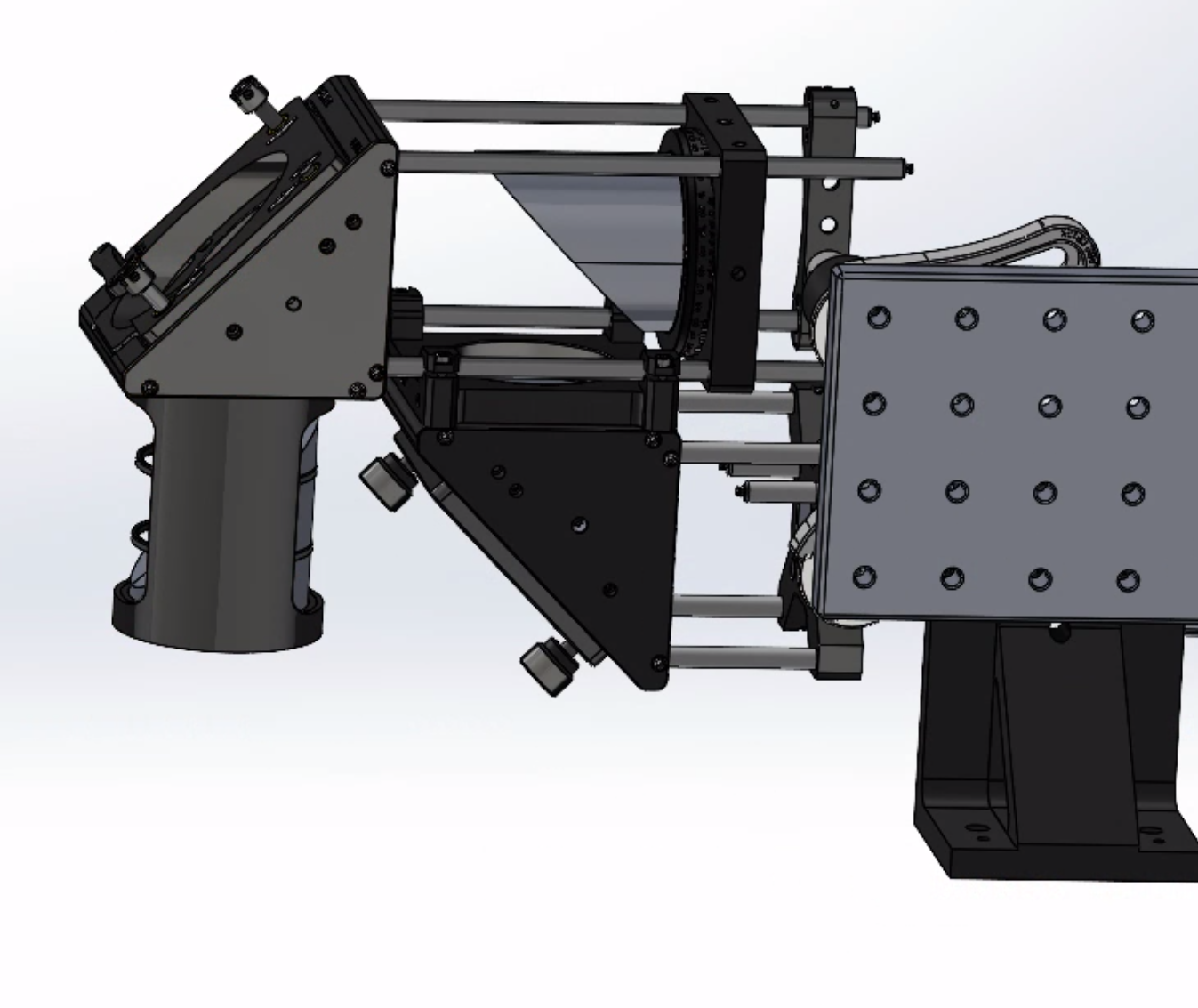
Sample Viewing System at ALS Beamline 12.3.2
Beamline 12.3.2 at the ALS is a microdiffraction facility. Its original sample viewing system was outdated, yielding low-quality images due to the stage’s tilting angle and long working distance. To address these issues, I developed a new microscope featuring a relay off-axis parabolic system and a tiltly compensated optics that provides an extended working distance and better sample viewing quality. This upgrade has significantly improved the imaging quality, ensuring more accurate specimen positioning.